SAQ 1
A 72-year-old male presents with central chest pain for 2 hours. The pain is sharp, radiates to his back, and appears to be diminishing. His observations are BP 210/100, HR 80, Sats 98% RA. There is no pulse or BP discrepancy between his arms. You suspect aortic dissection.
A 72-year-old male presents with central chest pain for 2 hours. The pain is sharp, radiates to his back, and appears to be diminishing. His observations are BP 210/100, HR 80, Sats 98% RA. There is no pulse or BP discrepancy between his arms. You suspect aortic dissection.
- List four risk factors for aortic dissection.
- HTN
- Age >60 years
- Male>Female (65% occur in males)
- Connective tissue disorders – Marfan’s, Ehler Danlos
- Cocaine use
- Previous cardiac surgery
- Arteritis
- Coarctation/aortic valve disease
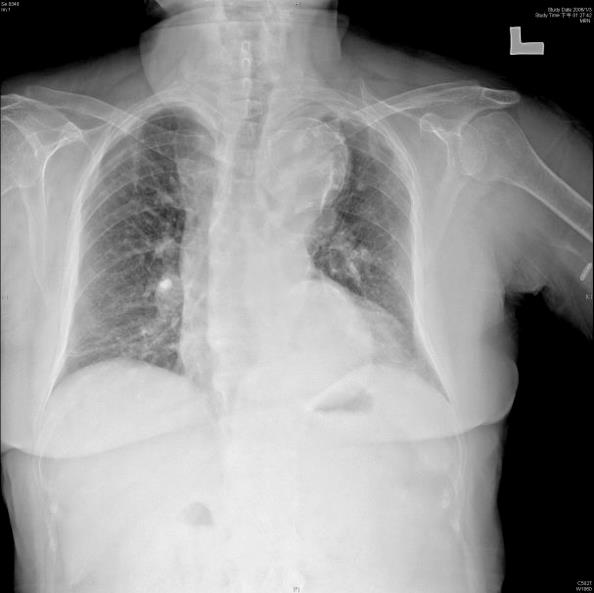
- The patient is moved to resus and a CXR obtained (below). List four features on this CXR suggestive of dissection.
- Displaced calcification
- Widened mediastinum
- Cardiomegaly
- Displaced trachea/LMB
- A CTA confirms a DeBakey type 3 dissection. List your management priorities for this dissection, giving endpoints where appropriate.
- No points for “surgical consult”
- Beta blocker – labetalol 10mg, esmolol 0.5 – 1mg, metoprolol IV 5mg
- BP control – IV SNP, GTN.
- Aim SBP 100-120 and HR 60-80
- Avoid reflex tachycardia
- List four contraindications to surgical repair of aortic dissection.
- Age >70
- Stroke symptoms
- Shock/hypotension
- MI
- Renal failure pre-op
- Previous AVR
- Bradycardia
SAQ 2
A 68-year-old Aboriginal female presents to your rural hospital with acute pain and paraesthesia in her right leg. Her medications include warfarin, digoxin, and metformin. She is a chronic smoker and has thrice weekly dialysis for end-stage renal failure.
- List three differential diagnoses for her presentation.
- Acute arterial occlusion (embolus from AF) – ischaemic limb
- DVT (phlegmasia cerulean dolens)
- Ischaemic intermittent claudication
- Buerger disease (thromboangiitis obliterans)
- Diabetic neuropathy
- Sciatica/spinal canal stenosis
- Arteritis
- Low output state (dialysis) superimposed on chronic arterial disease
- You suspect acute ischaemic limb. List four investigations you will order with justification.
- ECG – AF, MI
- Coags with INR – warfarin compliance, coag status
- CTA – identify location of obstruction
- CK – exclude rhabdomyolysis as complication of acute ischaemic limb
- Venous gas – lactate to allow monitoring of response to therapy, K+ level (renal patient)
- EUC etc
- CT angiography confirms acute 100% occlusion of the right femoral artery. What is the role of thrombolysis in this patient?
- Level 1a evidence – no evidence to support the use of thrombolysis in acute embolic limb ischaemia
SAQ 3
A 48-year-old male is yelling at triage, demanding urgent pain relief or he will leave. He is a “frequent flyer” to ED, with regular presentations related to alcohol abuse. He was seen two days prior after a fall and a below knee cast was applied for a fractured right tibia/fibula. He appears intoxicated.
- List three issues that need urgent management in this patient.
- Safety of staff – potential for violence – rapid de-escalation needed
- Safety of patient – may need care and control to keep him in ED
- Safety of limb – possible compartment syndrome – remove cast
- After calming the patient, you examine his right leg. The toes below the cast are white and swollen. List two clinical signs/symptoms strongly suggestive of an acute compartment syndrome.
- Pain out of proportion to clinical findings/examination
- Fascial compartment pressure > 30 mmHg
- Regarding the development of ischaemic limb:
- What is the traditional time frame after which ischaemia is likely to be irreversible?
- 6 hours
- What is the traditional time frame after which ischaemia is likely to be irreversible?
- Degree of muscle mass – more rapid onset of ischaemia with higher muscle mass
- What is the traditional time frame after which ischaemia is likely to be irreversible?
- Outline in order, the four steps you would take to manage his compartment syndrome in ED.
- Remove the cast (required to pass)
- Elevate limb to level of heart – not above, as will compromise arterial flow
- Analgesia/oxygen/IVT – avoid hypotension, this will worsen acidosis
- Fasciotomy if indicated – do all compartments of affected limb
- Seek and treat complications – rhabdomyolysis, metabolic acidosis, etc
- Urgent Ortho/Vasc input
SAQ 4
A 75-year-old male is brought to ED with 24 hours of melena. He has a past history of AAA with endoluminal stenting 10 months ago. His observations are BP 135/85, HR 80, GCS 15/15. On examination, he has a palpable non-tender aorta and melena on PR.
- Give three differential diagnoses for his presenting complaint.
- Aortenteric fistula (must include)
- Coagulapathy secondary to medication
- GIB
- You diagnose a likely aortoenteric fistula (AEF). What is the difference between a primary AEF and a secondary AEF?
- Primary: associated with AAA
- Secondary: associated with graft repair of a AAA
- List three methods of diagnosing an AEF in this patient with a pro and con for each method.
- Ultrasound at bedside – unlikely to see much, no radiation dose, repeatable
- CTA – high specificity/sensitivity, radiation dose
- Gastoduodenoscopy – can show other sources of GIB, but these don’t exclude AEF as two pathologies may co-exist
SAQ 5
A 23 year old painter recently diagnosed with Bordatella pertussis presents with gradual onset headache. The pain has been present for four days and is right sided. He states the pain came on after a particularly vigorous coughing episode.
- Give three differential diagnoses for his presentation.
- Carotid/vertebral artery dissection
- SAH
- Meningitis
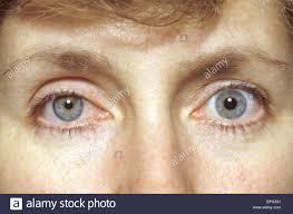
- On examination, you find the following (image). What is the likely diagnosis?
- Horner’s syndrome secondary to internal carotid artery dissection.
- List three management options for this condition, assuming the condition does not extend intracranially.
- Anticoagulation with heparin and warfarin (not strongly evidence based and validity unproven)
- Endovascular stenting
- Ligation of carotid artery/bypass procedure
SAQ 6
You are working as a senior registrar when an earthquake strikes the hospital you are working in, causing widespread damage to the building. Two hours into retrieval, you commence rescue of a young RMO who has been trapped by the left leg under masonry since the quake occurred.
- List two precautions you will take prior to attempting removal of the RMO from the masonry.
- Apply tourniquet to leg (controversial – UK recommends, Australia doesn’t, NZ does)
- Full monitoring and IV access
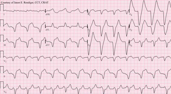
- Despite your best efforts, the RMO arrests soon after the leg is freed. An ECG is obtained during CPR (below). Briefly explain the pathophysiology behind this ECG.
- Crush syndrome – rhabdomyolysis – release of patient limb = release of large amounts K+, overwhelming hyperkalaemia = arrest.
- Outline how you will manage this electrolyte derangement acutely.
- Calcium chloride – 10ml of 10%
- Salbutamol 5mg neb
- Insulin 10 units actrapid IV with 50ml of 50% dextrose
- Sodium bicarbonate 50-100mmol
- Resonium
- What is the role of permissive hypotension in this trauma patient?
- Minimal – current recommendations are for mild fluid overload if tourniquet is applied, and mild hypotensive resuscitation if the pressure/tourniquet is released.

